Dr M.BARLA JOURNÉ – Orthopedist – Traumatologue – Surgery – Shoulder – Ankle – Pied – Rouen

Arthrose de cheville et de l’arrière-pied
Arthrose de la cheville
L’arthrose est l’usure ou la destruction du cartilage. Il n’y a actuellement aucun traitement qui permette de regénérer le cartilage. Les causes sont :
Idiopathique, c’est-à-dire que l’on ne retrouve pas de cause
Post traumatique (fractures)
Micro traumatique (entorses récidivantes de cheville)
En cas d’arthrose de cheville, les principales plaintes sont la douleur, le gonflement, les blocages. We also note a limitation of the walking perimeter and ankle mobility.. A weight-bearing radiographic assessment is essential.
Today there is 3 types of surgical treatment for ankle osteoarthritis :
Ankle arthroscopy
This treatment is carried out especially if the ankle osteoarthritis is not significant and the mobility of the joint remains normal.. The aim of the procedure is to clean the joint and remove foreign bodies.. Following the intervention, support is authorized with a walking boot 2 at 3 weeks. The effectiveness of this technique is almost immediate but remains transient. (some months). After a certain time, pain and other symptoms may recur.
Ankle arthrodesis
This is still the reference treatment today.. The principle is to fuse the ankle joint using screws or a plate., arthroscopically or openly. Following the surgery, support is prohibited during 4 at 6 weeks. The fusion time of the arthrodesis is 3 at 6 months depending on whether the surgery was done arthroscopically or openly. Arthrodesis makes it possible to turn a stiff and painful joint into a stiff and painless joint.. Following an ankle arthrodesis, walking is resumed without difficulty, there is almost no lameness.
Complications of surgery : pseudarthrodesis or failure of bone fusion (< 10 %) with a significantly increased risk in the event of tobacco poisoning, delayed skin healing if open surgery and if the patient smokes, hematoma, rare neurovascular lesion, algodystrophie.
Ankle prosthesis
It is indicated for patients who have painful ankle osteoarthritis but with retained mobility.. It is aimed at patients over 60 years. The main risk of this technique remains prosthetic loosening leading to subsequent arthrodesis..
Subtalar osteoarthritis
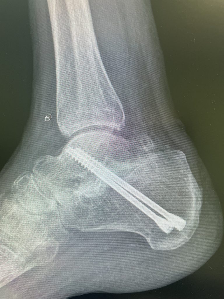
The subtalar joint is the joint located between the talus and the calcaneus., it is notably responsible for the movements purposes of the rearfoot and therefore the adaptation of our foot to steep or irregular terrain. The vast majority of subtalar osteoarthritis is post-traumatic (calcaneus fracture, fracture of the talus).
Treatment is very often surgical due to the poor tolerance of subtalar osteoarthritis..
Subtalar arthrodesis
This involves fusing the subtalar joint. The surgery is open or arthroscopic., the aim being to sharpen the subtalar articular surfaces then to carry out arthrodesis using 2 large caliber screw.
Hospitalization is 24-48 hours.
The recovery time is 6 month. Support is prohibited during 4 at 6 weeks.
Complications
Pseudarthrodesis especially if the patient smokes
Delayed skin healing if open surgery/smoking patient
Infection especially if smoking patient
Rare neurological lesion
Ankle tendinopathy
Tendinopathies d’Achille
The Achilles tendon is part of the suro-achilleo-plantar system (suro=triceps sural/muscle du mollet ; achilléo=tendon d’Achille ; plantar=plantar aponeurosis). Note that the Achilles tendon is the largest tendon in the body. He undergoes numerous constraints and develops tendinopathies.
Achilles tendinopathy causes weight-bearing but also inflammatory pain (at rest), we can see a thickening of the tendon or even redness. Palpation of the tendon is very painful. Sometimes, walking with a heel or heel is much better tolerated than walking with flat shoes.
Haglund's pathology is caused by repeated friction between the posterior-superior protuberance or hypertrophy of the calcaneus and the Achilles tendon.. There is often bursitis, but the Achilles tendon is often unharmed. This pathology is very often bilateral and mainly concerns young women..
Constraints in Achilles tendinopathy are common :
Repetitive microtrauma (running, trampling, prolonged standing, bad footwear)
Architectural Foot Disorders (hollow foot, Haglund's pathology, vertical calcaneus)
Gastrocnemius retraction (calf muscles)
Certain medications (fluoroquinolones)
We find 2 types of Achilles tendinopathy
- Corporal (damage to the body of the tendon)
- D’insertion (to its attachment to the calcaneus)
The imaging assessment includes a weight-bearing ankle x-ray, pour chercher des calcifications tendineuses ou des anomalies de position du calcanéum et une IRM qui va analyser précisément le tendon d’Achille.
Treatment is primarily medical, especially for corporal tendinopathy., because almost 75% of these tendinopathies are cured medically. Medical treatment includes : Plantar orthoses, weight loss/good hydration, rehabilitation based on stretching and eccentric work. Sometimes PRP injections are indicated. Infiltrations are not indicated due to the risk of tendon rupture.
Surgery for Achilles tendinopathy
In the event of failure of well-conducted medical treatment, the treatment will be surgical.
Lengthening of the gastrocnemius
If there is retraction of the gastrocnemius, the latter are elongated on the posterior surface of the knee. This gesture can be isolated or associated with other gestures. If the gesture is isolated, there is no immobilization and walking and rehabilitation are resumed immediately.
Corporal tendinopathy
We carry out arthroscopically, i.e. small skin incision and using a camera, debridement of the Achilles tendon to remove inflammation around it. Micro-incisions in the axis of the Achilles tendon (combing) can also be made. Hospitalization is outpatient. In the sequels, you are immobilized in a boot for 3 weeks with support authorized from the outset. Rehabilitation began at 3 weeks of surgery. The recovery time is 3 at 4 month.
Insertional tendinopathy
The surgery is performed open. The Achilles tendon must be detached from the calcaneus, remove bursitis, calcifications and the postero-superior protuberance of the calcaneus. The Achilles tendon is then reattached to the calcaneus via intraosseous anchors due to the high stresses exerted there..
You are immobilized in a boot without support for 4 weeks, rehabilitation begins at 1 month of intervention. The recovery time is 6 month. Resumption of sporting activities such as running, is done between 6 at 8 month post-operative.
Haglund's pathology
The surgery consists of arthroscopic removal of the postero-superior tuberosity of the calcaneus.. This surgery can also be performed openly. In case of arthroscopic excision, support is authorized from the outset under cover of a walking boot with heel pads during 4 weeks. En cas de chirurgie à ciel ouvert l’appui est interdit pendant 4 at 6 weeks. The recovery time is 3 month.
Ces chirurgies se font essentiellement en ambulatoire.
Complications
Retard de cicatrisation cutanée en cas de chirurgie à ciel ouvert et surtout si le patient est tabagique
Infection
Hématome
Œdème de cheville
Récidive rare
Raideur de cheville, algodystrophie
Ankle tendinopathy (Achille excepté)
Sont retrouvés essentiellement 3 types de tendinopathies.
Tendinopathie des fibulaires
Elle fait suite à une entorse de la cheville et s’accompagne d’œdème en arrière de la malléole externe, pain when mobilizing the ankle. A clinical and imaging assessment is essential to detect a dislocation of the peroneal tendons., cracking or tenosynovitis, i.e. effusion in the fibular sheath.
The primary treatment is conservative : plantar orthoses made, rehabilitation for muscle strengthening and proprioception work, weightloss. In case of failure of medical surgical treatment, surgical treatment is proposed : tendon release and debridement arthroscopically, i.e. with small skin incisions and a camera. Support is authorized from the outset with a walking boot during 3 weeks. Rehabilitation began at 3 weeks of surgery. The recovery time is 3 month. Resumption of sporting activities can be done at 4 month post-operative.
Posterior tibial tendon tendinopathy
The posterior tibial tendon is very important because it supports the medial arch of the foot. It is constantly stressed during support and prevents the foot from collapsing.
Posterior tibial tendinopathy follows trauma, to overweight, to hyper demands, medial instability of the ankle or an abnormality of the architecture of the foot (flat foot). It causes internal ankle pain upon mobilization and palpation., swelling under the medial malleolus. These pains are essentially in support. Ultrasound and MRI allow for precise diagnosis (tenosynovitis, tendinosis or tendon rupture). Treatment is primarily medical : weightloss, foot orthosis and rehabilitation.
In the event of well-conducted medical treatment in the event of failure of medical treatment, surgery may be offered. If the posterior tibial tendon is still present and inflamed, arthroscopic tendon debridement is proposed. If the treatment if the posterior tibial tendon is ruptured in the context of decompensated flat foot, depending on the lesions on clinical examination, more complex surgery is proposed : tendon transfer, osteotomy or arthrodesis. Support is authorized from the outset with a walking boot during 3 weeks. Rehabilitation begins three weeks after the operation. The recovery time is 4 at 6 month.
Flexor hallucis longus tendinopathy
The causes of flexor hallucis longus tendinopathy are quite similar to those of posterior tibial tendon tendinopathy.. The failure of medical treatment raises the question of surgical treatment by arthroscopic tendon release and debridement.. The recovery time is 3 at 4 month, support resumed immediately under cover of a walking boot which should be kept 3 weeks.
Complications :
Edema, hematoma, infrequent failure, ankle stiffness, algodystrophie.
Chronic ankle instability
Recurrent sprains/ankle instability
Ankle sprains are extremely common. 10% trauma arriving in an emergency department is secondary to ankle sprains.
In the majority of cases, These ankle sprains involve the external lateral ligament (breaking or stretching). Le traitement en aigu est le plus souvent fonctionnel selon le protocole RICE.
Rest : repos / Ice : icing / Contention : immobilization with splint but with preservation of support / Elévation : on elevation of the injured limb to reduce edema. Sometimes immobilization with a cast without support is essential.
Rehabilitation must be implemented fairly quickly from D10-15 after the trauma.
There is little room for acute surgical treatment.
But the complaints (pain, recurrence of sprains) are numerous (10-45%) as well as the after-effects (20%) : chronic lateral ankle instability progresses to ankle osteoarthritis with cartilage destruction.
L’instabilité latérale de cheville est multifactorielle :
- External lateral ligament incompetence
- Muscle deficit
- Hyperlaxité
- Architectural Foot Disorders (varus, hollow foot)
Surgery is indicated, in the event of persistent lateral instability, despite well-conducted rehabilitation.
We are using 2 types of surgical treatment, qui ont an efficiency of 85-95% on stability :
- Ligament Repair
- Anatomic Ligamentoplasty of the Ankle
Surgery for ankle instability
Ligament repair
This involves repairing and re-tensioning the ruptured external ligament by fixing it via an anchor on the external malleolus.. This procedure is done under arthroscopy, that is to say with a camera and with small incisions. Support is immediate and is done under cover of a walking boot for 1 months with start of rehabilitation at 1 month.
Hospitalization is outpatient.
The recovery time is 2 at 3 month.
This surgery is indicated in young and athletic patients., in the event of isolated lesions of the anterior external ligament and if the trauma is less than 4 month.
Complications :
Infection rare, hematoma, edema, rare recurrence.
Anatomical ligamentoplasty of the ankle
This involves reconstructing the entire lateral ankle ligament using a hamstring tendon (the gracilis) which is taken below the knee. The technique is called anatomical because we reconstruct the external ligament using its paths..
Hospitalization is often outpatient.
Support is immediate, under cover of a walking boot while 3 weeks. Rehabilitation begins immediately. Sports activities are resumed between 2 at 4 month post-operative. The recovery time is 3 at 6 month.
Complications :
Infection rare, hematoma, edema, rare recurrence, douleurs résiduelles.

Le docteur Manuela Barla-Journé chirurgienne orthopédiste, traite les problèmes musculosquelettiques de l’adulte. Elle s’occupe plus généralement des traumatismes et des maladies de l’appareil locomoteur : tendons, ligaments, os, muscles et articulations. Elle est référent pour traiter les problèmes articulaires de l’épaule, de la cheville et du pied. Le docteur Manuela Barla-Journé ne traite pas les ongles incarnés. Le docteur Manuela Barla-Journé ne prend pas en charge les patients de moins de 15 years.
https://www.icos-normandie.com/manuela-barlajourne/
MAKE AN APPOINTMENT
#orthopedist #orthopedist #traumatology #traumatologist #specialist #trauma #traumatologyoffoot #traumatologyoflepaule #traumatologyofacheville #interventionchirurgicale #specialistentraumatology #chirurgiedelepaule #chirurgiedupied #chirurgiedelacheville #chirurgienrouen #chirurgie #epaule #ankle #foot #rouen #normandie #eure

